Cancer is one of the leading causes of morbidity and mortality around the world. Despite some success, traditional anticancer drugs developed to reduce tumor growth face important limitations primarily due to undesirable bone marrow and cardiovascular toxicity. Many drugs fail in clinical development after showing promise in preclinical trials, suggesting that the available in vitro and animal models are poor predictors of drug efficacy and toxicity in humans. Thus, novel models that more accurately mimic the biology of human organs are necessary for high-throughput drug screening. Three-dimensional (3D) microphysiological systems can utilize induced pluripotent stem cell technology, tissue engineering, and microfabrication techniques to develop tissue models of human tumors, cardiac muscle, and bone marrow on the order of 1 mm(3) in size. A functional network of human capillaries and microvessels to overcome diffusion limitations in nutrient delivery and waste removal can also nourish the 3D microphysiological tissues. Importantly, the 3D microphysiological tissues are grown on optically clear platforms that offer non-invasive and non-destructive image acquisition with subcellular resolution in real time. Such systems offer a new paradigm for high-throughput drug screening and will significantly improve the efficiency of identifying new drugs for cancer treatment that minimize cardiac and bone marrow toxicity.
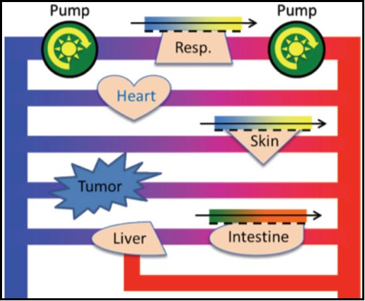
A strategy for integrating essential three-dimensional microphysiological systems of human organs for realistic anticancer drug screening